5005 S. Cooper St.
Suite 250
Arlington, TX 76017
Best Kidney Cancer Doctors in Dallas-Fort Worth
Kidney Cancer Is Scary
No one wants to hear, “You have kidney cancer.” About 75,000 people in the United States are diagnosed with the disease during their lifetime. Knowing you’re not alone doesn’t make it easier. We understand how overwhelming the worry and flood of information can be. That’s why we help men and women understand their kidney cancer and all their treatment options. Empowered with knowledge, they can take control and choose the best course of action for them.

There’s Encouraging News!
Understanding the “T” word.
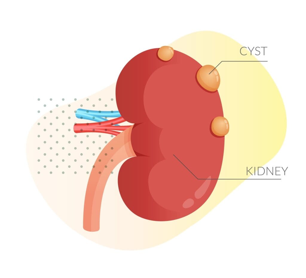
The term “tumor” refers to an area in an organ that is not typically supposed to be there. A tumor may also be referred to as a “mass,” “lesion,” “neoplasm” or “cyst.” All of these terms mean the same thing, but none of them indicates whether or not a tumor isa cancer. Often, tumors are found accidentally when an individual is undergoing X-rays for other reasons. Sometimes, they are found when investigating why a patient is experiencing side pain or blood in the urine.
How do we know when a tumor is cancer?
Simple Cysts (Bosniak I) are “simple” water-filled bubbles in the kidney. They do not have any internal structure. They are typically perfectly round. They can be very small (1/8 inch) or they can be very large (up to about 8 inches). They are typically not associated with any pain or symptoms. They are not cancers and they do not turn into cancers. They almost never need any kind of treatment or surgery. They are sometimes monitored with additional imaging tests to see if they are changing or growing over time. Rarely, the fluid might be drained from the cyst. This is typically done by a radiologist under light anesthesia. This procedure might be performed to tell whether the cyst is causing pain. If a simple cyst is thought to be causing pain or blockage of the urinary tract, laparoscopic surgery may be recommended to remove the top of the cyst. This is called laparoscopic cyst decortication.
Hemorrhagic/Proteinaceous Cysts are a specific type of “complex” cyst that contain either blood or a thicker protein fluid inside. These cystic masses are not cancers and do not require surgery. They do typically need to be watched with repeated imaging tests.
Complex Cysts (Bosniak II, III, IV) are cyst-type masses that have some internal tissue or structure. This category of cysts is named after the radiologist who first suggested classifying them into different types—Dr. Morton Bosniak. The Bosniak type is typically decided by the radiologist and urologist after reviewing a CT or MRI with contrast injected into a vein. The Bosniak types are:
Bosniak II and IIF Cysts Bosniak II and IIF Cysts are the least complex of all the “complex” cysts. This type of cyst may contain some calcium and have internal walls called septations. While the septations do not have measurable blood flow inside them, the cyst walls may be a little thicker than the walls of a “simple” cyst. Almost always benign (non-cancerous), they rarely require surgical removal, but may be monitored with additional imaging tests over time.
Bosniak III Cysts Bosniak III Cysts have several internal features that separate these cystic masses from “simple” or other “complex” cysts. They can have thicker walls and some of the walls may show blood flow inside them on the imaging test. As many as half of these can be cancer. These cysts can sometimes be observed with additional imaging tests, although many of them will need to be removed surgically. If the cyst is removed and found to be cancer, almost all patients are cured and do not need any additional treatment.
Bosniak IV Cysts Bosniak IV Cysts are the most complex type of cystic masses. These lesions typically have a solid portion that has blood flow inside it. Most of these lesions are removed surgically, and determined to be cancer after they are removed. After surgery, almost all patients are cured and do not need any additional treatment.
Health Navigator
- Harrison “Mitch” Abrahams, MD
- Jeffrey Charles Applewhite, MD
- Jerry Barker, MD, DABR, FACR
- Paul Benson, MD
- Richard Bevan-Thomas, MD
- Keith D. Bloom, MD
- Tracy Cannon-Smith, MD, FMS
- Paul Chan, MD
- Kara Choate, MD
- Lira Chowdhury, DO, FACOS
- Weber Chuang, MD
- Adam Cole, MD, FS
- M. Patrick Collini, MD
- Zachary Compton, MD
- Adam Hollander, MD
- Patrick A. Huddleston, MD
- Justin Tabor Lee, MD
- Wendy Leng, MD, FPMRS
- Alexander Mackay, MD
- Tony Mammen, MD
- F.H. “Trey” Moore, MD
- Geofrey Nuss, MD
- Christoper Pace, MD
- Jason Poteet, MD
- Andrew Y. Sun, MD
- Scott Thurman, MD
- James Clifton Vestal, MD, FACS
- Keith Waguespack, MD
- Diane C. West, MD
- Keith Xavier, MD, FPMRS
Related News & Information
Life After the Lift (UroLift) is Great!
For many men, an enlarged prostate can make life miserable. The urgent need to go, urinating more frequently than normal, a weak urinary stream, difficulty starting and stopping—they’re all symptoms of BPH. Find out how the UroLift is helping men get their life back.
There Are Effective Treatments for Kidney Tumors
Appropriate treatment is determined by the size and composition of the tumor. The physician may recommend one of the following treatment options.

Kidney Tumor Biopsy
A needle biopsy may be done to see what the cells of the mass look like under a microscope and determine if the mass is cancer. A biopsy is typically performed by an interventional radiologist and patients are given a light anesthetic. Afterward, there may be mild pain or blood in the urine, but patients usually recover quite quickly. Patients would need to be off blood-thinning medications before and after this procedure. Biopsy results usually take a week or so to come back.
Kidney Cyst Drainage
If your doctor is concerned that your “simple” kidney cyst is causing pain or blocking the urine tube (ureter), a cyst drainage procedure may be recommended. Typically, this is done by an interventional radiologist and under light anesthetic. There may be mild pain after the procedure, but typically patients recover quite quickly. Blood-thinning medications should not be used before or after this procedure.


Robotic and Laparoscopic Surgery
Urology Partners uses several types of robotic and laparoscopic procedures to treat kidney tumors and masses.
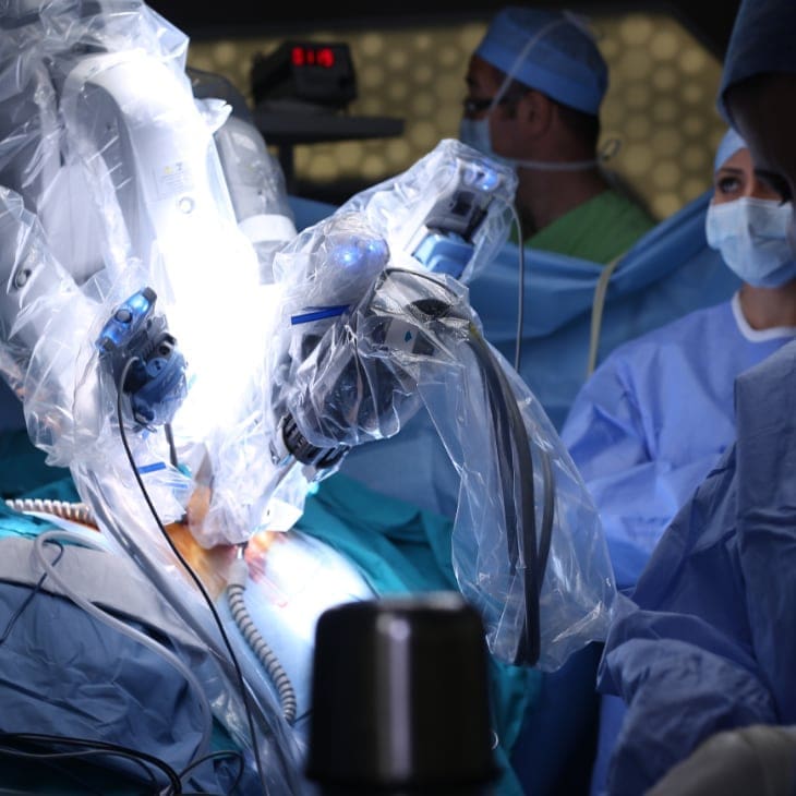
Also known as partial kidney removal, this is the most common procedure for kidney tumors that need to be removed. The physicians at UPNT were some of the first doctors in the world to do this procedure in 2004, and have successfully performed several thousands of these operations. The technique involves using the robotic da Vinci Surgical System to safely remove the kidney tumor and sew the kidney back together with stitches. This treatment option is mainly used for smaller kidney tumors. Patients must be off blood-thinning medications before and after this procedure.
During this surgical procedure, patients are under complete anesthesia. Once the mass is removed, it is sent to a pathologist to determine if it is cancer. Results for the pathology test usually take about one week. Patients spend one or two nights in the hospital after this surgery, and are prescribed pain medicine and IV fluids. Most patients recover from this operation within two to three weeks. A post-op appointment with your UPNT the surgeon is scheduled to ensure healing is taking place as it should.
The advantage of robotic surgery over traditional open or laparoscopic surgery is that the surgeon can see much better, has better control of the instruments and patients recover more quickly with less pain.
Also known as complete kidney removal, this is the second most common surgery for kidney masses. During the robotic version of this surgery, several keyhole incisions are made to telescopically remove the kidney. With laparoscopic radical nephrectomy, a slightly larger incision is made to remove the kidney. It is typically used for larger kidney masses, masses located deeper in the kidney, masses that might invade surrounding areas, and for kidneys that no longer function.
During this surgical procedure, patients are under complete anesthesia. Once the mass is removed, it is sent to a pathologist to determine if it is cancer. Results for the pathology test usually take about one week. Patients typically spend one or two nights in the hospital after this surgery, and are prescribed pain medicine and IV fluids. Most patients recover from this operation within two to three weeks. A post-op appointment with your UPNT surgeon is scheduled to ensure healing is taking place as it should.
The advantage of robotic surgery over traditional open surgery is that the surgeon can see much better and patients recover more quickly with less pain.
This minimally invasive surgery(cryoablation) destroys tumor cells by freezing them. This procedure is done through tiny keyhole incisions using telescopes and ultrasound to find the mass. The tumor is frozen to well below -20° Celsius. It is then allowed to defrost slightly, then frozen a second time to ensure the cells are dead. At this very cold temperature, the dead cells turn into scar tissue.
Patients must be off blood-thinning medications before and after this procedure. Often, the surgeon will biopsy the mass immediately before treating it. Once the mass is removed, it is sent to a pathologist to determine if it is cancer. Results for the pathology test usually take about one week. Patients typically spend one night in the hospital after this surgery, and are prescribed pain medicine and IV fluids. Most patients recover from this operation within two to three weeks. A post-op appointment with the surgeon is scheduled to ensure healing is taking place as it should.
This surgery involves removing the diseased kidney and the entire kidney tube (ureter) that connects the tube to the bladder. This surgery is typically recommended for patients who have cancer or tumors of the urinary tract (kidney or ureter tube).
During this surgical procedure, patients are under complete anesthesia. Once the mass is removed, it is sent to a pathologist to determine if it is cancer. Results for the pathology test usually take about one week. Patients typically spend one or two nights in the hospital after this surgery, and are prescribed pain medicine and IV fluids. Many patients are sent home with a catheter to drain the bladder, which helps the bladder heal. Most patients recover from this operation within two to three weeks. A post-op appointment with the surgeon is scheduled to ensure healing is taking place as it should.
The advantage of this surgery over traditional open surgery is that the surgeon can see much better and the patients recover more quickly with less pain.
This minimally invasive surgery is done to remove a “simple” (Bosniak I) cyst that is either causing pain or blocking the flow of urine to the bladder. Patients must be off blood-thinning medications before and after this procedure. Patients are under complete anesthesia during the procedure, typically spend one night in the hospital after surgery, and are prescribed pain medicine and IV fluids. Most patients recover within two to three weeks. A post-op appointment with the surgeon is scheduled to ensure healing is taking place as it should.
The advantage of this procedure over traditional open surgery is that the surgeon can see much better and the patients recover more quickly with less pain.
We Are Your Kidney Cancer Experts
Call for an Appointment

Two-Time Kidney Cancer Survivor Donald Kyle’s Message to African-American Men
Don Kyle’s father was just 56 when he died from cancer. He thinks about that—a lot. “As an African-American male, I know that my father had a reluctance to go to doctors,” Kyle says. “I don’t know why, our family had good healthcare. I know it’s something that is endemic in the African- American community.

